The Interdepartmental Equipment Facility
Gel Imaging and Photography

Proteins and nucleic acids are frequently labeled with a radioisotope such as 32P, to facilitate their identification and quantitation following chromatographic separation. Techniques based on the use of radioactive labels are still widely used in applications that require high sensitivity. Quantification of results is facilitated by exposure of the sample to autoradiography film, or to a reusable storage phosphor screen (IP). The latent image of the spot distribution on the gel is next developed by an Imager such as the FLA-5000. On the other hand, due to the need for safe handling and disposal of hazardous waste, the use of radioactive tracers is frequently limited to manual, low-throughput protocols.

In recent years, fluorescence, and chemiluminescence have emerged as alternative technologies to the traditional radioisotope-based systems. Significant advances in fluorescent and luminescence dye chemistry were accompanied by the development of advanced imaging systems with extensive data processing capabilities. Convenience, speed and safety as well as sensitivity and resolution are important factors in deciding which label to use.
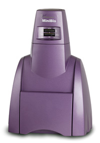
Our Gel-Imaging facility offers imaging instruments from both categories, which are listed below. The DNR Minibis is an imaging system for fluorescence and the CLBIS was designed to monitor and image Enhanced Chemiluminescence or ECL. On the other hand we have the Agfa Curix X-ray film Developer for radiolabeled samples, and the Fujifilm FLA-5000 which can carry out imaging of both fluorescent gels and of imaging plates previously exposed to radioactive samples.
List of Available Equipment
| Instrument | Make & Model |
| Gel Documentation System | DNR Minibis Pro |
| Luminescence Imaging System | DNR CLBIS |
| Radioisotope and Fluorescence Imager | Fuji FLA-5000 |
| X-ray Film Processor | Agfa Curix 60 |